Head CT: brain and bone windowsWhat is the diagnosis?
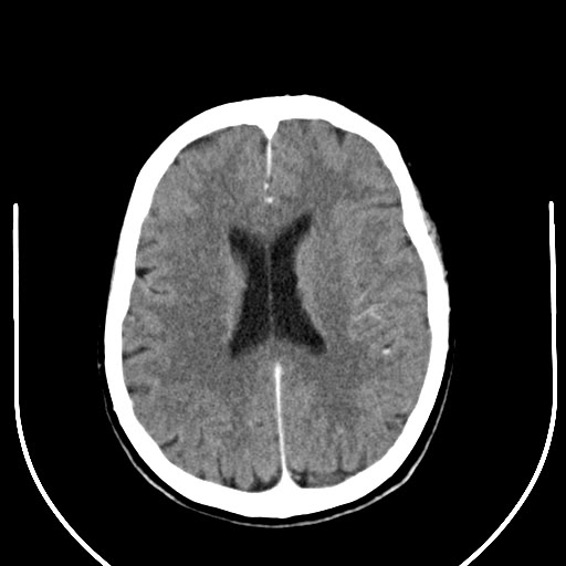
CT head without contrast
4.8 mm brain kernel (H21s) · 1.2 mm brain kernel (H21s) · 1.2 mm bone kernel (H70h)
Scroll with the mouse wheel. Right-drag (or the contrast tool) changes window width and level; try the CT presets: brain, subdural, bone.
Images: National Cancer Institute CPTAC, CPTAC-CM collection, via The Cancer Imaging Archive, CC BY 4.0 — data citation. Every slice at full depth; 3D works best on a laptop or desktop.
 CT
1 / 2
Axial 4.8 mm slice, brain window (W 80 / L 40).
Image: National Cancer Institute CPTAC, CPTAC-CM collection, via The Cancer Imaging Archive, CC BY 4.0 — sourceAnswer hidden. Review the images and history, decide on your diagnosis, then reveal.
Findings
Three axial series of the same scan are provided: a 4.8 mm reconstruction for a quick read, and thin 1.2 mm reconstructions with a soft-tissue (brain) kernel and a sharp (bone) kernel.
- In the brain window (W 80 / L 40) grey and white matter separate, the ventricles and sulci are easy to judge, and fresh blood would stand out as bright.
- In the bone window (W 2800 / L 600) the brain turns uniform grey but the calvarium, skull base, mastoid air cells and paranasal sinuses become sharp. This is where fractures are found.
- The bone kernel looks grainy in a brain window: sharp kernels trade noise for edge detail.
This study comes from a public research archive and has not been formally reported by a Radflow radiologist. It is here to teach viewing technique and anatomy, not to establish a diagnosis.
Case discussion
Why windowing matters. CT stores density in Hounsfield units (air −1000, water 0, brain about 35, fresh blood 60–80, bone several hundred and above). A screen shows about 256 grey levels, so a window chooses which slice of that range is spread from black to white. Reading every head CT in at least a brain, a subdural (W 200 / L 80) and a bone window avoids the classic misses: a thin subdural haematoma against the skull, and a non-displaced fracture.
Kernels are not windows. The kernel is chosen when the images are reconstructed from the raw data; the window is chosen when you look at them. A bone kernel cannot be recreated by windowing a soft-tissue series.
Why no 3D here? This scan was acquired step-and-shoot, so the slice spacing jumps where the blocks join. The viewer therefore treats it as a stack, not a volume. For multiplanar and 3D reconstruction see the next case, a helical scan with even 1 mm spacing.
More cases like this.
All cases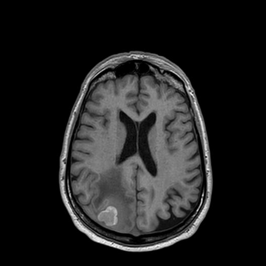 3D 3
Multiple melanoma brain metastases on MRI
Brain MRI of an adult with cutaneous melanoma, from a research cohort. A full routine protocol is available: T1, T2, FLAIR, DWI/ADC, gradient echo and 3D T1 before and after contrast.
 3D 2
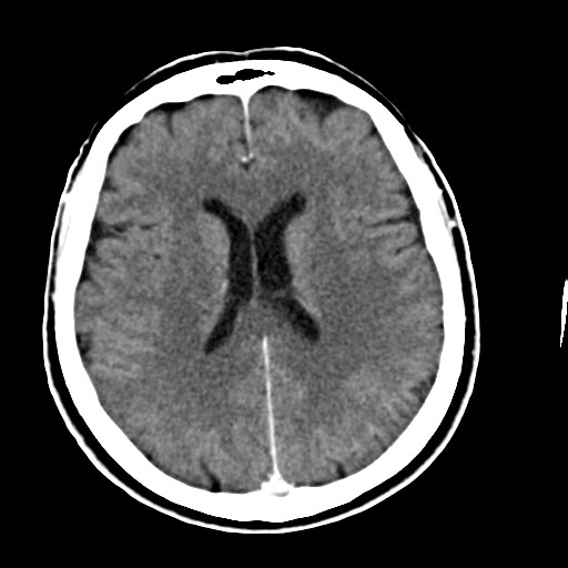
Head CT with contrast: MPR and 3D reconstruction
Contrast-enhanced CT head of an adult from a melanoma research cohort, reconstructed at 1 mm. Use it to practise multiplanar and 3D viewing.
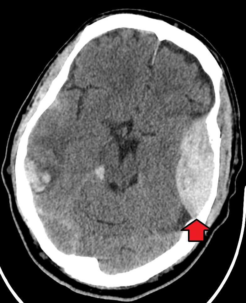
Extradural hematoma
Head injury after a fall. Brief loss of consciousness, now increasingly drowsy.