Head CT with contrast: MPR and 3D reconstructionWhat is the diagnosis?
CT head with contrast
1 mm soft-tissue kernel · 1 mm bone kernel · 5 mm axial
Open “MPR + 3D” for linked axial, coronal and sagittal views with a 3D volume rendering. In the 3D view pick a bone preset and drag to rotate.
Images: National Cancer Institute CPTAC, CPTAC-CM collection, via The Cancer Imaging Archive, CC BY 4.0 — data citation. Every slice at full depth; 3D works best on a laptop or desktop.
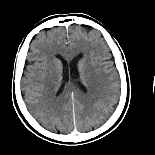 CT
1 / 2
Axial 1 mm slice after contrast, brain window.
Image: National Cancer Institute CPTAC, CPTAC-CM collection, via The Cancer Imaging Archive, CC BY 4.0 — sourceAnswer hidden. Review the images and history, decide on your diagnosis, then reveal.
Findings
A helical acquisition with evenly spaced 1 mm slices forms a true volume. From it the viewer builds:
- Multiplanar reconstruction (MPR): coronal and sagittal planes, linked by crosshairs, at the same resolution as the axial images. The planes can be tilted (oblique MPR) to line up with the skull base, as on a scanner console.
- Slab views: thicken a plane into a maximum-intensity projection (MIP) to follow enhancing vessels, or an average (AIP) to smooth noise.
- 3D volume rendering: each voxel gets a colour and opacity from its density. A bone preset makes soft tissue transparent and shows the skull; a soft-tissue preset shows the skin surface.
This study comes from a public research archive and has not been formally reported by a Radflow radiologist. It is here to teach viewing technique and anatomy, not to establish a diagnosis.
Case discussion
What makes a series reconstructable. MPR and 3D need thin slices (ideally 1.25 mm or less), constant spacing and the same orientation for every slice. Thick 5 mm slices still reformat, but coronal and sagittal images look stepped.
Where 3D helps. Complex skull and facial fractures, craniosynostosis, surgical planning, and CT angiography (aneurysms, vessel anatomy). For everyday diagnosis, thin axial slices and MPR remain the reference; 3D is a map, not a replacement.
Volumetrics. The same volume lets you segment a structure, for example a haematoma, and read its volume in mL from the voxel count. That is more accurate than the bedside ABC/2 estimate.
More cases like this.
All cases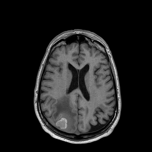 3D 3
Multiple melanoma brain metastases on MRI
Brain MRI of an adult with cutaneous melanoma, from a research cohort. A full routine protocol is available: T1, T2, FLAIR, DWI/ADC, gradient echo and 3D T1 before and after contrast.
 DICOM 2
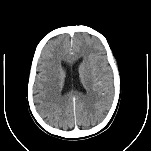
Head CT: brain and bone windows
Non-contrast CT head of an adult from a melanoma research cohort. Use it to practise windowing and to compare soft-tissue and bone reconstructions.
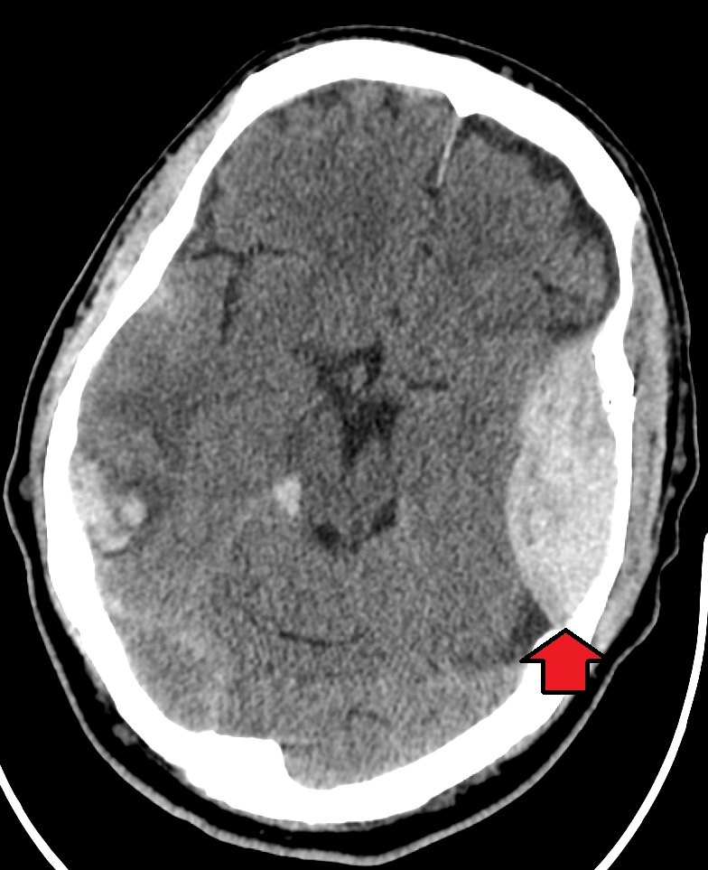
Extradural hematoma
Head injury after a fall. Brief loss of consciousness, now increasingly drowsy.