Multiple melanoma brain metastases on MRIWhat is the diagnosis?
MRI brain, routine protocol with contrast
Sagittal T1 · axial T2 · axial FLAIR · DWI b1000 and ADC · coronal GRE · 3D T1 pre and post contrast
Choose a 2×2 layout and drag different sequences into each panel to compare them. MPR works on the 3D T1 series only.
Images: National Cancer Institute CPTAC, CPTAC-CM collection, via The Cancer Imaging Archive, CC BY 4.0 — data citation. Every slice at full depth; 3D works best on a laptop or desktop.
 MRI
1 / 3
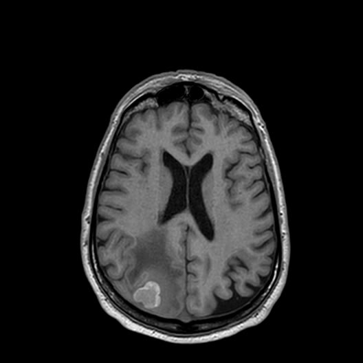
Axial 3D T1 before contrast: a high-signal lesion in the right parietal lobe.
Image: National Cancer Institute CPTAC, CPTAC-CM collection, via The Cancer Imaging Archive, CC BY 4.0 — sourceAnswer hidden. Review the images and history, decide on your diagnosis, then reveal.
Findings
- Several intra-axial lesions. The most conspicuous is in the right parietal lobe, with further lesions in the left hemisphere.
- The lesions are bright on the T1 images obtained before contrast, a feature typical of melanoma metastases (melanin and/or blood products).
- Surrounding vasogenic oedema, high signal on FLAIR and T2, follows the white matter.
- Compare the post-contrast T1, the DWI/ADC pair and the gradient-echo series to judge enhancement, diffusion and haemorrhage.
Interpretation prepared by Radflow from the images and the cohort’s diagnosis of cutaneous melanoma, and reviewed by the Radflow team; it is not the original radiology report.
Case discussion
MRI is read as a set of sequences, not one image. There are no Hounsfield units: signal intensity depends on the sequence, so each series is judged against its own normal pattern (fluid dark on T1 and FLAIR, bright on T2). A viewer for MRI must let you hang several series side by side and scroll them together.
Melanoma metastases. Melanoma is one of the cancers most likely to spread to the brain. Metastases are typically multiple, sit at the grey–white junction, enhance after contrast and carry disproportionate oedema. Intrinsic T1 hyperintensity, from melanin or haemorrhage, is a helpful clue, and gradient-echo or SWI shows blood products as dark blooming.
Differential for multiple enhancing lesions: metastases, abscesses (marked restricted diffusion centrally), multifocal glioma, demyelination (incomplete ring) and lymphoma.
More cases like this.
All cases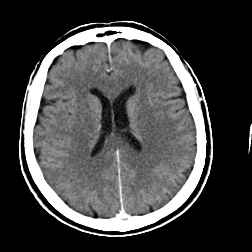 3D 2
Head CT with contrast: MPR and 3D reconstruction
Contrast-enhanced CT head of an adult from a melanoma research cohort, reconstructed at 1 mm. Use it to practise multiplanar and 3D viewing.
 DICOM 2
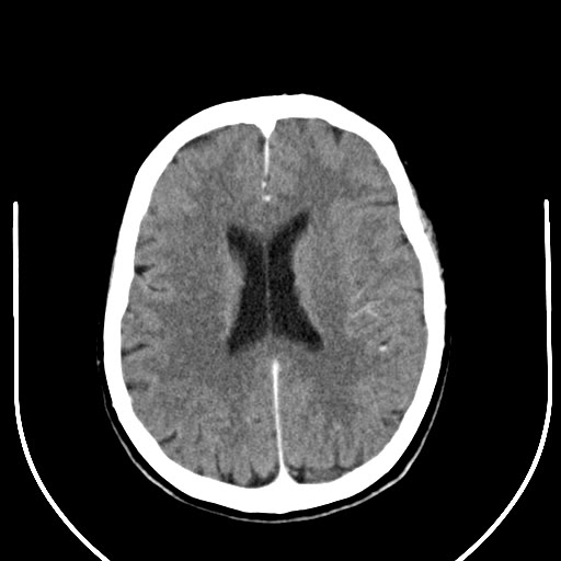
Head CT: brain and bone windows
Non-contrast CT head of an adult from a melanoma research cohort. Use it to practise windowing and to compare soft-tissue and bone reconstructions.
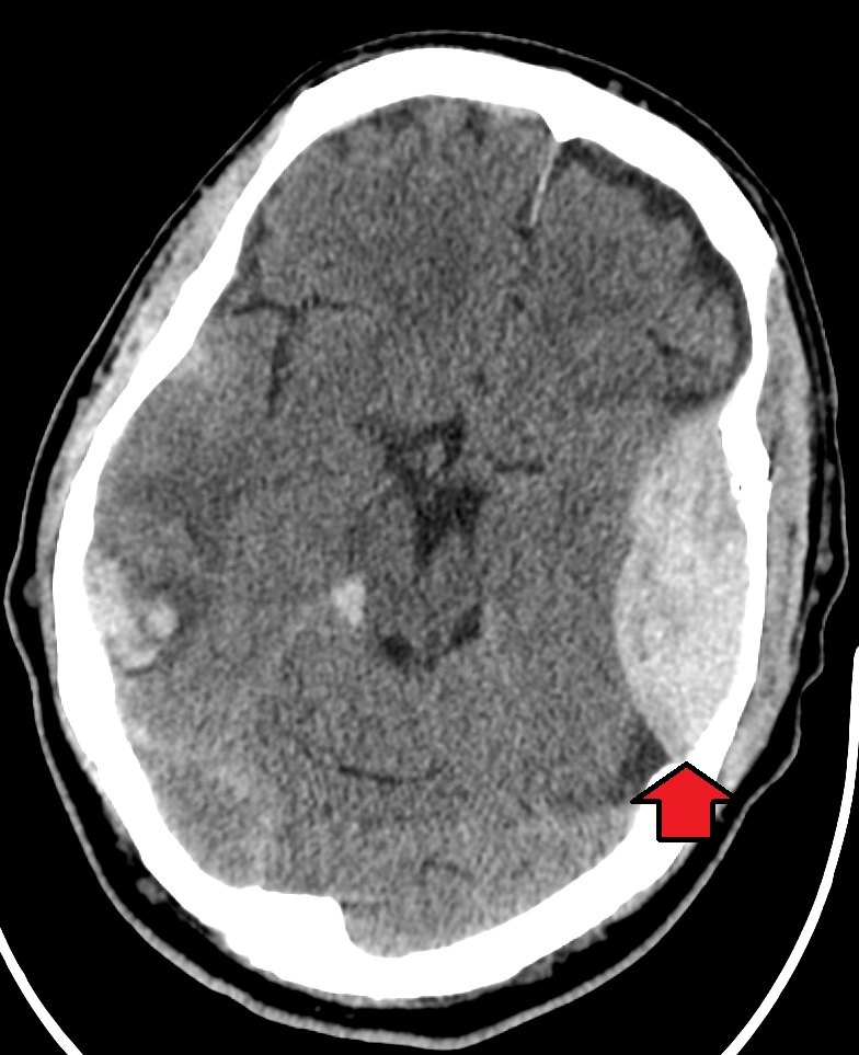
Extradural hematoma
Head injury after a fall. Brief loss of consciousness, now increasingly drowsy.