Case 1003
Right lower zone pneumoniaWhat is the diagnosis?
 X-ray
1 / 1
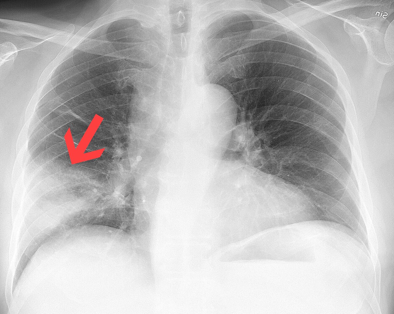
Frontal chest radiograph with an arrow from the original source.
Image: Mikael Häggström, M.D., CC0 — sourceAnswer hidden. Review the images and history, decide on your diagnosis, then reveal.
Findings
- Airspace opacity in the right mid to lower zone (arrow)
- The right heart border remains visible, favouring the right lower lobe over the middle lobe
- No significant volume loss; costophrenic angles clear
- Left lung clear
Case discussion
Airspace consolidation in a patient with fever and productive cough is most likely pneumonia. Because the right heart border is preserved, the opacity is probably posterior, in the lower lobe — a lateral view would confirm this.
Consolidation keeps lung volume, unlike collapse. Radiographic resolution lags behind clinical recovery; a follow-up film at about six weeks is often advised in smokers and older adults to exclude an underlying lesion.