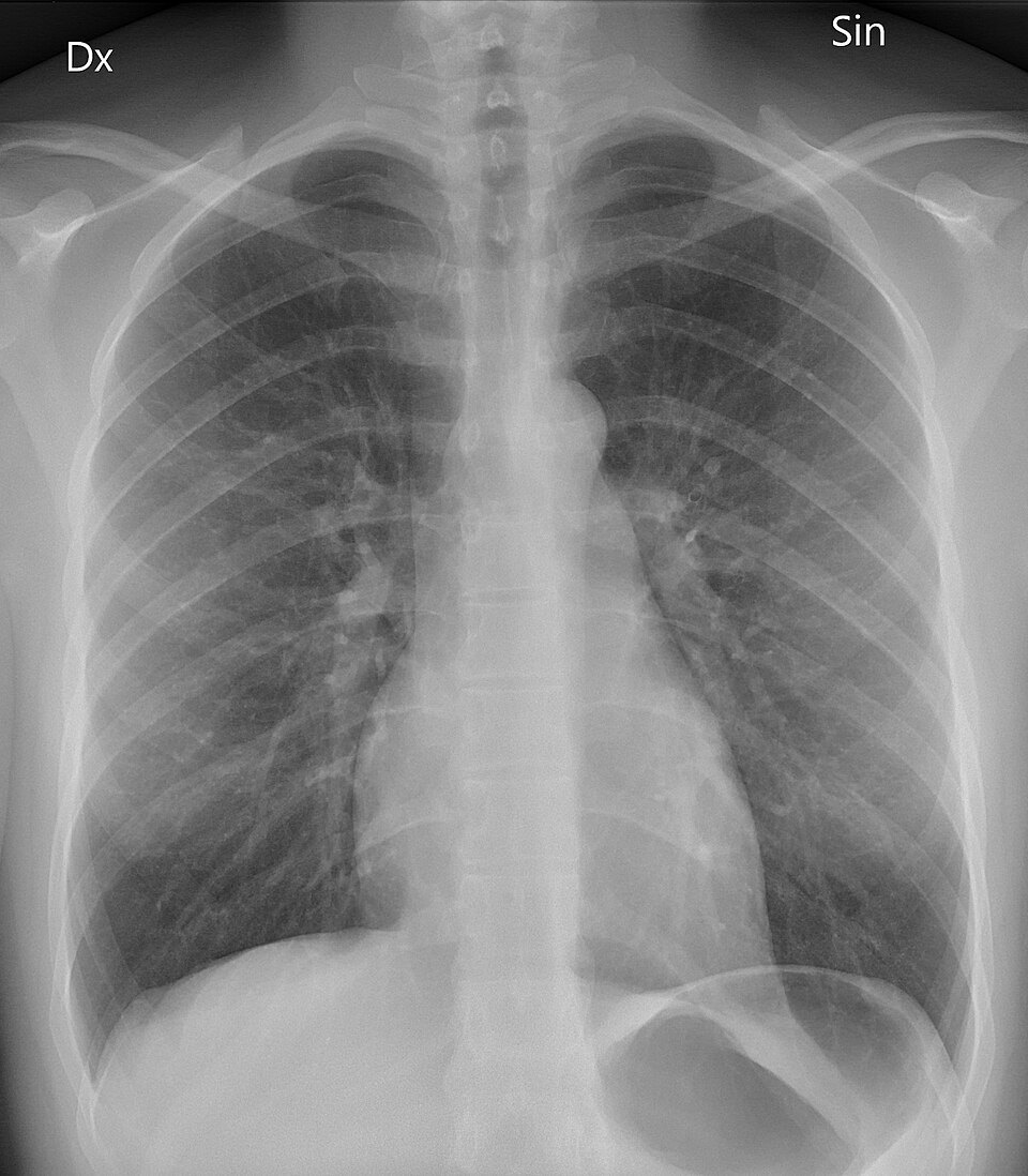
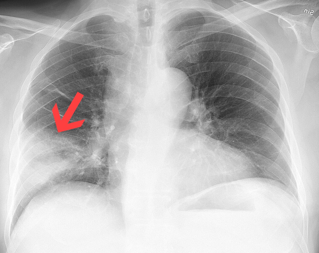
The silhouette sign describes the loss of a normally visible interface — such as a heart border or hemidiaphragm — when the lung directly next to it becomes airless. Two structures of the same density in contact cannot be told apart on a radiograph.
Radiographic features
The sign localises disease on a single frontal film:
- Right heart border lost: right middle lobe
- Left heart border lost: lingula
- Hemidiaphragm lost: lower lobe
- Aortic knuckle lost: left upper lobe (apicoposterior segment)
- Right paratracheal stripe lost: right upper lobe
Conversely, if a border remains crisp despite an overlying opacity, the abnormality lies in a different plane — for example a lower lobe process behind a preserved heart border.
Practical points
A poorly inspired or rotated film can blur borders and mimic the sign. Check technique before calling it.
Cases and figures
References
- Felson B, Felson H. Localization of intrathoracic lesions by means of the postero-anterior roentgenogram; the silhouette sign. Radiology. 1950;55(3):363–374.
Cite this entry
Silhouette sign. Radflow Encyclopedia. Last revised 12 November 2026. https://radflow.cc/encyclopedia/silhouette-sign (accessed 15 November 2026).
Related entries
Educational reference for healthcare professionals and students. It is not medical advice and does not replace clinical judgement or local guidelines.