Case 1001
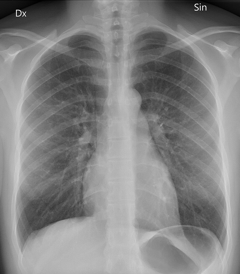
Normal chest radiographWhat is the diagnosis?
 X-ray
1 / 1
Frontal (PA) chest radiograph.
Image: Mikael Häggström, CC0 — sourceAnswer hidden. Review the images and history, decide on your diagnosis, then reveal.
Findings
Well-centred, adequately inspired PA radiograph.
- Trachea central; normal carina and hila
- Lungs clear, with lung markings reaching the chest wall on both sides
- Heart size normal (cardiothoracic ratio under 0.5); sharp heart borders
- Clear costophrenic angles; normal hemidiaphragms with the right slightly higher than the left
- No bony or soft tissue abnormality
Case discussion
Knowing the normal is the foundation of reading any film. Use this case to practise a systematic search: check technique (rotation, inspiration, projection, exposure), then airway, lungs, heart, diaphragm and the review areas — apices, behind the heart, below the diaphragm, hila and bones.
Notice how every border is crisp. When a structure next to the heart or diaphragm becomes airless, that border disappears — the silhouette sign.
Encyclopedia
Cardiothoracic ratioThe maximum horizontal width of the heart divided by the maximum internal width of the thorax on a PA chest radiograph.Chest radiograph: systematic reviewA structured way to read every chest radiograph, from technical quality to review areas, so that subtle findings are not missed.Silhouette signLoss of a normal border between soft tissue and air when an adjacent structure fills with fluid or tissue, used to localise chest disease.