Quiz — what’s the diagnosis?
Look at the images, read the history and pick the most likely diagnosis. Your score is kept on this device.
0Correct
0Answered
–Score
 X-ray
1 / 1
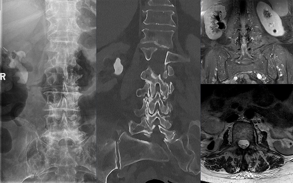
Composite: plain radiograph, coronal CT and MRI of the same patient.
Image: Hellerhoff, CC BY-SA 4.0 — source
Case 1013
Skip this case
Presentation
Recurrent urinary tract infections. Imaged during work-up.
- A large, branching radiopaque calculus projects over the right kidney on the plain film
- Coronal CT confirms a dense stone filling the right renal pelvis and extending into the calyces
- On MRI the stone is a signal void within the collecting system