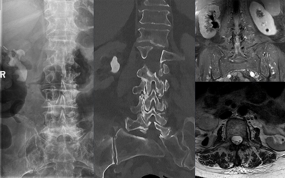
Urolithiasis is the formation of stones in the urinary tract. Calcium oxalate stones are the most common. A staghorn calculus branches to fill the renal pelvis and calyces and is often associated with infection by urease-producing bacteria (struvite).
Clinical presentation
Ureteric colic: severe loin-to-groin pain with haematuria, nausea and restlessness. Fever with an obstructed kidney is a urological emergency. Staghorn stones may cause recurrent infection with little pain.
Radiographic features
Non-contrast CT (CT KUB)
- Nearly all stones are hyperdense and visible
- Secondary signs: hydronephrosis, perinephric stranding, ureteric dilatation
- The soft tissue rim sign distinguishes a ureteric stone from a phlebolith
Plain radiograph
Most calcium stones are radiopaque. A KUB film is useful for follow-up of known opaque stones.
Ultrasound
Shows hydronephrosis and larger renal stones without radiation; a good first test in pregnancy and children.
Treatment and prognosis
Small ureteric stones often pass with analgesia and medical expulsive therapy. Larger or obstructing stones need ureteroscopy, shockwave lithotripsy or, for staghorn calculi, percutaneous nephrolithotomy. Infected obstructed systems need urgent drainage.
Cases and figures
References
- Smith-Bindman R, Aubin C, Bailitz J, et al. Ultrasonography versus computed tomography for suspected nephrolithiasis. N Engl J Med. 2014;371(12):1100–1110.
- Türk C, Petřík A, Sarica K, et al. EAU guidelines on diagnosis and conservative management of urolithiasis. Eur Urol. 2016;69(3):468–474.
Cite this entry
Urolithiasis. Radflow Encyclopedia. Last revised 2 November 2026. https://radflow.cc/encyclopedia/urolithiasis (accessed 15 November 2026).
Educational reference for healthcare professionals and students. It is not medical advice and does not replace clinical judgement or local guidelines.