Pulmonary embolism (PE) occurs when thrombus, most often from a deep vein thrombosis, lodges in the pulmonary arterial tree. Risk factors include immobility, surgery, cancer, pregnancy, oestrogen therapy and inherited thrombophilia.
Clinical presentation
Breathlessness, pleuritic chest pain, haemoptysis, tachycardia, hypoxia or collapse. Clinical probability scores (Wells, Geneva) combined with D-dimer decide who needs imaging.
Radiographic features
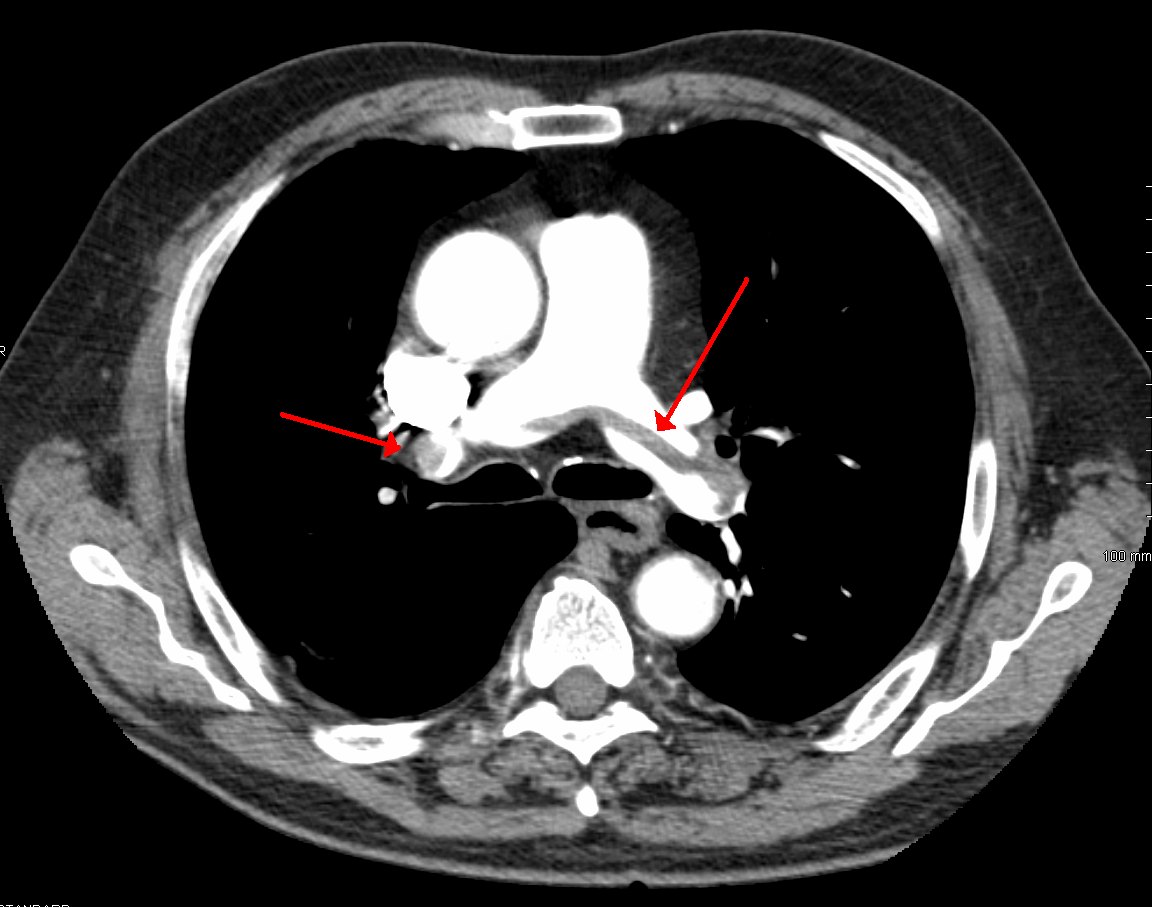
CT pulmonary angiography
- Filling defects within contrast-opacified pulmonary arteries
- A saddle embolus straddles the bifurcation of the main pulmonary artery
- Right heart strain: right ventricle larger than the left, septal bowing, reflux of contrast into the IVC
- Peripheral wedge-shaped infarcts
Chest radiograph
Often normal; it is mainly used to exclude other causes. Rare signs include Hampton hump and Westermark sign.
V/Q scintigraphy
An alternative when contrast is contraindicated and in pregnancy.
Treatment and prognosis
Anticoagulation for most patients. Thrombolysis is reserved for high-risk PE with haemodynamic instability; catheter-directed or surgical options exist for selected cases.
Practical points
Poor contrast timing and breathing artefact can mimic emboli. Check that filling defects are seen on more than one slice and plane.
Cases and figures
References
- Konstantinides SV, Meyer G, Becattini C, et al. 2019 ESC Guidelines for the diagnosis and management of acute pulmonary embolism developed in collaboration with the European Respiratory Society. Eur Heart J. 2020;41(4):543–603.
Cite this entry
Pulmonary embolism. Radflow Encyclopedia. Last revised 4 November 2026. https://radflow.cc/encyclopedia/pulmonary-embolism (accessed 15 November 2026).
Related entries
Educational reference for healthcare professionals and students. It is not medical advice and does not replace clinical judgement or local guidelines.