Aortic dissection begins with an intimal tear through which blood tracks into the media. The Stanford classification guides management: type A involves the ascending aorta, type B does not.
Clinical presentation
Abrupt, tearing chest or back pain, often maximal at onset. Pulse or blood pressure differences between arms, a new aortic regurgitation murmur, stroke, limb ischaemia or abdominal pain reflect branch involvement. Hypertension and connective tissue disease are major risk factors.
Radiographic features
CT angiography
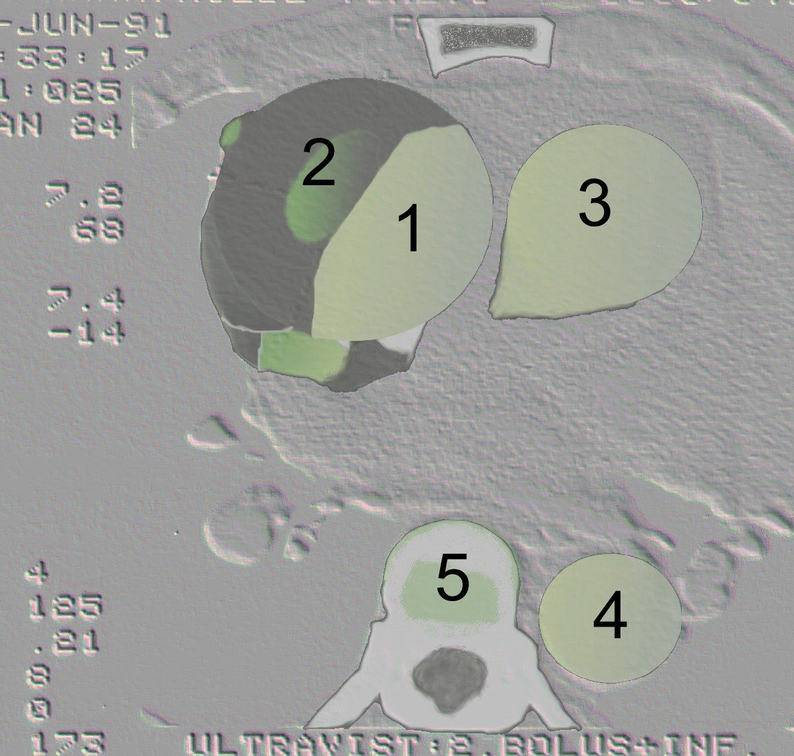
- An intimal flap separating true and false lumens
- The false lumen is usually larger and enhances later
- Extension into the arch vessels, coronary, mesenteric, renal or iliac arteries
- Complications: pericardial haemopericardium, rupture, malperfusion
Chest radiograph
May show mediastinal widening or an abnormal aortic contour, but a normal film does not exclude dissection.
Echocardiography
Transoesophageal echo is useful in unstable patients and shows aortic regurgitation and pericardial fluid.
Treatment and prognosis
Type A dissection is a surgical emergency. Uncomplicated type B is managed with blood pressure and heart rate control; complicated type B is treated with endovascular stenting.
Differential diagnosis
- Intramural hematoma
- Penetrating atherosclerotic ulcer
- Motion artefact in the ascending aorta
Cases and figures
References
- Erbel R, Aboyans V, Boileau C, et al. 2014 ESC Guidelines on the diagnosis and treatment of aortic diseases. Eur Heart J. 2014;35(41):2873–2926.
Cite this entry
Aortic dissection. Radflow Encyclopedia. Last revised 3 November 2026. https://radflow.cc/encyclopedia/aortic-dissection (accessed 15 November 2026).
Related entries
Educational reference for healthcare professionals and students. It is not medical advice and does not replace clinical judgement or local guidelines.