Pneumoperitoneum is free gas within the peritoneal cavity. Outside the post-operative period it usually means perforation of a hollow viscus — most often a peptic ulcer or a colonic diverticulum.
Clinical presentation
Sudden severe abdominal pain, peritonism and a rigid abdomen. Elderly or immunosuppressed patients and those on steroids may have surprisingly few signs.
Radiographic features
Erect chest radiograph
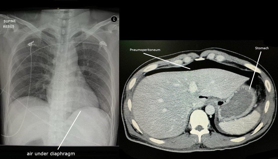
- A crescent of lucency under one or both hemidiaphragms
- The patient should be upright for several minutes beforehand so gas can rise
- Beware Chilaiditi sign (bowel between liver and diaphragm), which shows haustral folds
Supine abdominal radiograph
- Rigler sign: both sides of the bowel wall are visible
- Falciform ligament sign and the football sign in large collections
CT
The most sensitive test: even a few bubbles of free gas are seen anteriorly on lung windows. CT often shows the site of perforation.
Treatment and prognosis
Resuscitation, antibiotics and urgent surgical review. A small, contained leak may occasionally be managed conservatively.
Practical points
Gas may persist for several days after laparotomy or laparoscopy, so correlate with the surgical history.
Cases and figures
References
- Stapakis JC, Thickman D. Diagnosis of pneumoperitoneum: abdominal CT vs. upright chest film. J Comput Assist Tomogr. 1992;16(5):713–716.
Cite this entry
Pneumoperitoneum. Radflow Encyclopedia. Last revised 10 November 2026. https://radflow.cc/encyclopedia/pneumoperitoneum (accessed 15 November 2026).
Related entries
Educational reference for healthcare professionals and students. It is not medical advice and does not replace clinical judgement or local guidelines.