PneumoperitoneumWhat is the diagnosis?
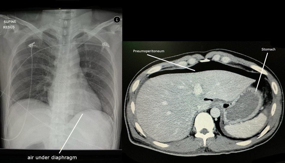 X-ray
1 / 2
Supine chest radiograph and axial CT, annotated in the original source.
Image: Cerevisae, CC BY-SA 4.0 — sourceAnswer hidden. Review the images and history, decide on your diagnosis, then reveal.
Findings
- Lucency beneath the diaphragm, separating it from the underlying viscera
- On CT, free gas collects anteriorly in front of the liver and stomach
- The abdominal film in the second patient also contains abnormal free gas outlining bowel and peritoneal structures
Case discussion
Free intraperitoneal gas without recent surgery means a perforated hollow viscus until proved otherwise. An erect chest radiograph is the classic first test, but CT is far more sensitive and often shows the cause.
On supine films, look for Rigler's sign (both sides of the bowel wall visible), a visible falciform ligament and the football sign. Beware mimics such as Chilaiditi sign, where interposed colon shows haustral folds.
Encyclopedia
More cases like this.
All cases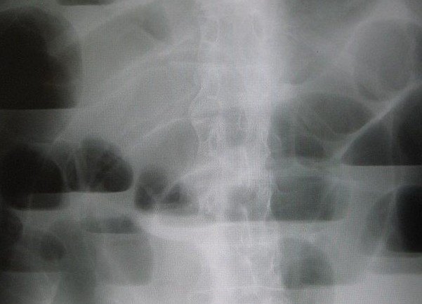 2
Small bowel obstruction
Colicky central abdominal pain, vomiting and distension. Previous laparotomy.
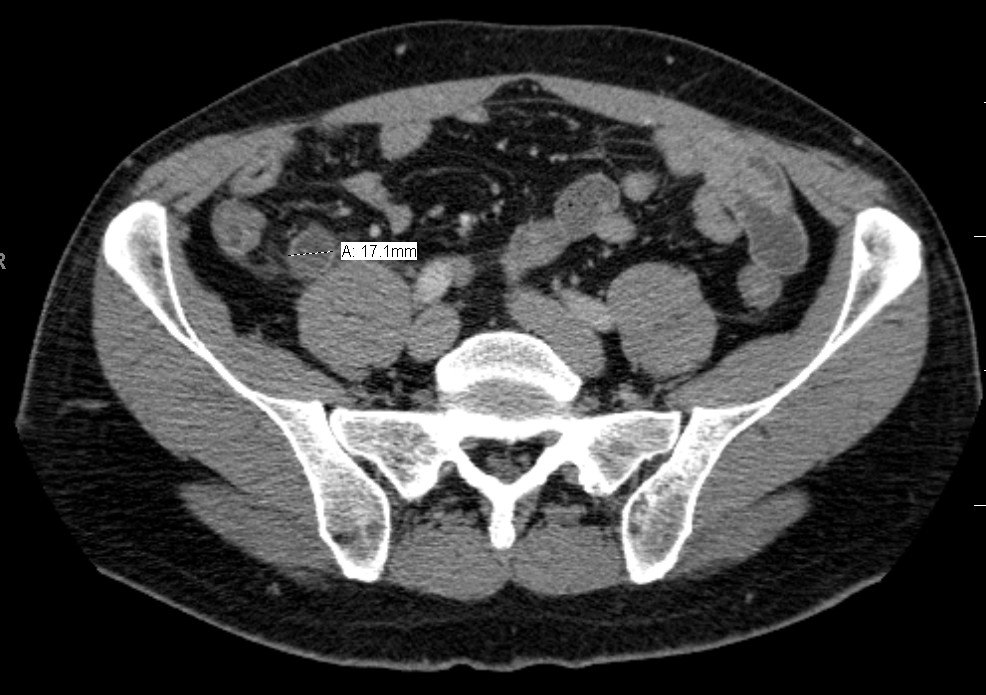 2
Acute appendicitis
Periumbilical pain migrating to the right iliac fossa, with anorexia and low-grade fever.
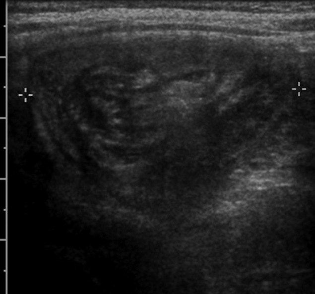
Ileocolic intussusception
Episodes of inconsolable crying with drawing up of the legs, and vomiting.