An extradural (epidural) hematoma is blood between the inner table of the skull and the dura. Around 90% are associated with a skull fracture, most often tearing the middle meningeal artery in the temporal region.
Clinical presentation
The classic lucid interval — brief loss of consciousness, recovery, then rapid deterioration — occurs in only a minority. Headache, vomiting, falling GCS, an ipsilateral fixed dilated pupil and contralateral weakness signal mass effect.
Radiographic features
CT
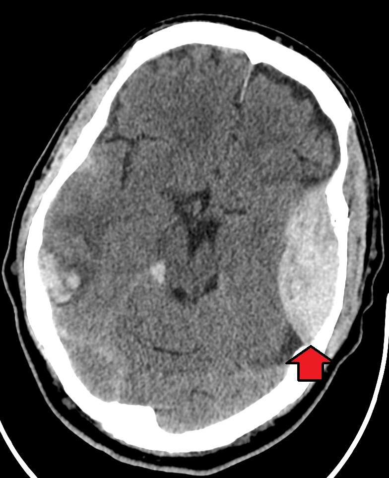
- Biconvex (lentiform) hyperdense collection
- Limited by cranial sutures, where the dura is firmly attached, but can cross the midline and the tentorium
- Mixed density (the swirl sign) suggests active bleeding
- Overlying fracture on bone windows
- Mass effect: effacement of sulci, midline shift, herniation
Treatment and prognosis
Large collections, significant midline shift or neurological decline need urgent craniotomy. When evacuated promptly, outcomes are often excellent because the underlying brain may be largely uninjured.
Differential diagnosis
- Acute subdural hematoma (crescentic, crosses sutures, does not cross the midline)
- Dural-based tumour
Practical points
Always review bone windows in head trauma; a subtle temporal fracture is a clue to an evolving extradural bleed.
Cases and figures
References
- Bullock MR, Chesnut R, Ghajar J, et al. Surgical management of acute epidural hematomas. Neurosurgery. 2006;58(3 Suppl):S7–S15.
Cite this entry
Extradural hematoma. Radflow Encyclopedia. Last revised 6 November 2026. https://radflow.cc/encyclopedia/extradural-hematoma (accessed 15 November 2026).
Related entries
Educational reference for healthcare professionals and students. It is not medical advice and does not replace clinical judgement or local guidelines.