Cholelithiasis means stones in the gallbladder. Most are cholesterol or mixed stones; pigment stones occur with haemolysis and cirrhosis. Many people never develop symptoms.
Clinical presentation
Biliary colic: episodic right upper quadrant or epigastric pain after meals, lasting hours. Complications include acute cholecystitis, choledocholithiasis with obstructive jaundice or cholangitis, gallstone pancreatitis and, rarely, gallstone ileus.
Radiographic features
Ultrasound
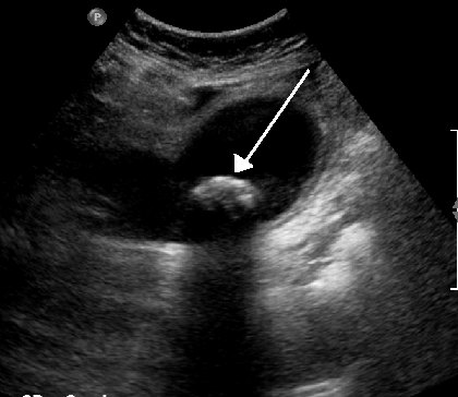
- Echogenic foci within the gallbladder lumen
- Posterior acoustic shadowing
- Mobility with change of patient position
- A contracted gallbladder full of stones gives the wall–echo–shadow sign
Signs of acute cholecystitis include wall thickening over 3 mm, pericholecystic fluid and a positive sonographic Murphy sign.
CT and MRI
Many stones are invisible on CT because they match bile density. MRCP is the best non-invasive test for stones in the common bile duct.
Treatment and prognosis
Asymptomatic stones are usually left alone. Symptomatic gallstones are treated with laparoscopic cholecystectomy; bile duct stones are removed by ERCP or at surgery.
Differential diagnosis
- Gallbladder polyp (non-shadowing, non-mobile)
- Biliary sludge
- Gas in the adjacent duodenum
Cases and figures
References
- National Institute for Health and Care Excellence. Gallstone disease: diagnosis and management (CG188). London: NICE; 2014.
Cite this entry
Cholelithiasis. Radflow Encyclopedia. Last revised 7 November 2026. https://radflow.cc/encyclopedia/cholelithiasis (accessed 15 November 2026).
Educational reference for healthcare professionals and students. It is not medical advice and does not replace clinical judgement or local guidelines.