Case 1006
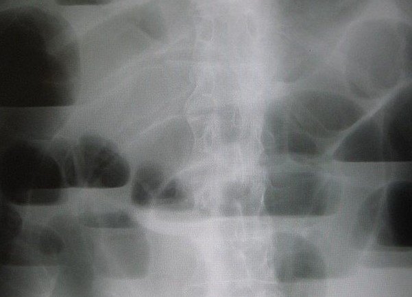
Small bowel obstructionWhat is the diagnosis?
Answer hidden. Review the images and history, decide on your diagnosis, then reveal.
Findings
- Multiple dilated, centrally located small bowel loops
- Numerous air–fluid levels at different heights on the erect film
- CT shows dilated, fluid-filled small bowel loops with air–fluid levels
Case discussion
Dilated small bowel with multiple air–fluid levels in a patient with previous surgery suggests adhesional small bowel obstruction.
CT confirms the diagnosis, finds the transition point and — most importantly — looks for strangulation: reduced wall enhancement, mesenteric oedema, free fluid or a closed loop. Those features mean surgery; otherwise most adhesional obstructions settle with decompression and fluids.
Encyclopedia
More cases like this.
All cases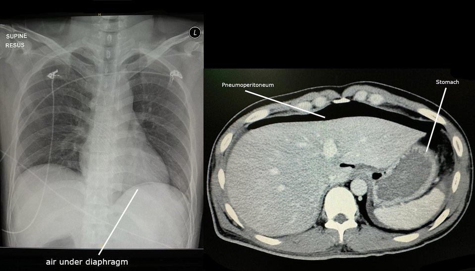 2
Pneumoperitoneum
Sudden severe epigastric pain, now generalised. Rigid abdomen on examination.
 2
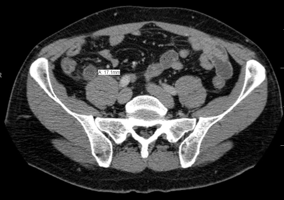
Acute appendicitis
Periumbilical pain migrating to the right iliac fossa, with anorexia and low-grade fever.
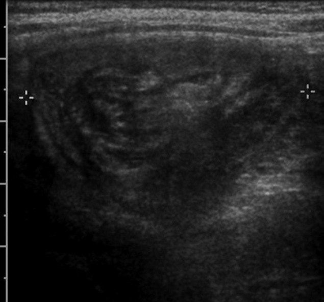
Ileocolic intussusception
Episodes of inconsolable crying with drawing up of the legs, and vomiting.